
Page 63 - Revista Portuguesa - SPORL - Vol 52 Nº3
P. 63
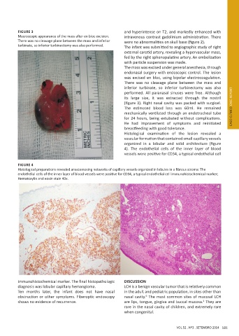
FIGURE 3                                          and hyperintense on T2, and markedly enhanced with
          Macroscopic appearance of the mass after en bloc excision.   intravenous contrast gadolinium administration. There
          There was no cleavage plane between the mass and inferior   were no abnormalities on skull base (figure 2).
          turbinate, so inferior turbinectomy was also performed.   The infant was submitted to angiographic study of right
                                                            external carotid artery, revealing a hypervascular mass,
                                                            fed by the right sphenopalatine artery. An embolization
                                                            with particle suspension was made.
                                                            The mass was excised under general anesthesia, through
                                                            endonasal surgery with endoscopic control. The lesion
                                                            was excised en bloc, using bipolar electrocoagulation.
                                                            There was no cleavage plane between the mass and
                                                            inferior turbinate, so inferior turbinectomy was also
                                                            performed. All paranasal sinuses were free. Although
                                                            its large size, it was extracted through the nostril
                                                            (figure 3). Right nasal cavity was packed with surgicel.
                                                            The  estimated  blood  loss  was  60ml.  He  remained       CASO CLÍNICO CASE REPORT
                                                            mechanically ventilated through an endotracheal tube
                                                            for  24  hours,  being  extubated  without  complications.
                                                            He  had  improvement  of  symptoms  and  reinitiated
                                                            breastfeeding with good tolerance.
                                                            Histological  examination  of  the  lesion  revealed  a
                                                            vascular formation that contained small capillary vessels
                                                            organized  in  a  lobular  and  solid  architecture  (figure
                                                            4). The endothelial cells of the inner layer of blood
                                                            vessels were positive for CD34, a typical endothelial cell

          FIGURE 4
          Histological preparations revealed anastomosing networks of capillary vessels organized in lobules in a fibrous stroma: The
          endothelial cells of the inner layer of blood vessels were positive for CD34, a typical endothelial cell immunohistochemical marker;
          Hematoxylin and eosin stain 40x.


          immunohistochemical marker. The final histopathologic   DISCUSSION
          diagnosis was lobular capillary hemangioma.       LCH is a benign vascular tumor that is relatively common
          Ten months later, the infant does not have nasal   in the adult and pediatric population, in sites other than
          obstruction or other symptoms. Fiberoptic endoscopy   nasal cavity.  The most common sites of mucosal LCH
                                                                      6
          shows no evidence of recurrence.                  are lips, tongue, gingiva and buccal mucosa.  They are
                                                                                                 2
                                                            rare in the nasal cavity of children, and extremely rare
                                                            when congenital.


                                                                                     VOL 52 . Nº3 . SETEMBRO 2014 181