
Page 62 - Revista Portuguesa - SPORL - Vol 52 Nº3
P. 62
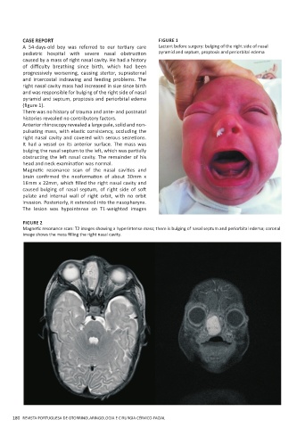
CASE REPORT                                        FIGURE 1
         A  54-days-old  boy  was  referred  to  our  tertiary  care   Lactent before surgery: bulging of the right side of nasal
         pediatric  hospital  with  severe  nasal  obstruction   pyramid and septum, proptosis and periorbital edema
         caused by a mass of right nasal cavity. He had a history
         of  difficulty  breathing  since  birth,  which  had  been
         progressively worsening, causing stertor, suprasternal
         and intercostal indrawing and feeding problems. The
         right nasal cavity mass had increased in size since birth
         and was responsible for bulging of the right side of nasal
         pyramid and septum, proptosis and periorbital edema
         (figure 1).
         There was no history of trauma and ante- and postnatal
         histories revealed no contributory factors.
         Anterior rhinoscopy revealed a large pale, solid and non-
         pulsating mass, with elastic consistency, occluding the
         right nasal cavity and covered with serous secretions.
         It had a vessel on its anterior surface. The mass was
         bulging the nasal septum to the left, which was partially
         obstructing the left nasal cavity. The remainder of his
         head and neck examination was normal.
         Magnetic  resonance  scan  of  the  nasal  cavities  and
         brain  confirmed  the  neoformation  of  about  30mm  x
         16mm x 22mm, which filled the right nasal cavity and
         caused  bulging  of  nasal  septum,  of  right  side  of  soft
         palate and internal wall of right orbit, with no orbit
         invasion. Posteriorly, it extended into the nasopharynx.
         The lesion was hypointense on T1-weighted images

          FIGURE 2
          Magnetic resonance scan: T2 images showing a hyperintense mass; there is bulging of nasal septum and periorbital edema; coronal
          image shows the mass filling the right nasal cavity.


      180  REVISTA PORTUGUESA DE OTORRINOLARINGOLOGIA E CIRURGIA CÉRVICO-FACIAL