
Page 68 - Revista Portuguesa - SPORL - Vol 62. Nº1
P. 68
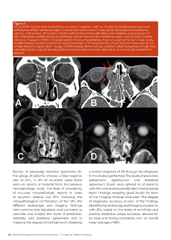
Figure 3
          CT and MRI studies with and without contrast in a patient with an IP with intraorbItal extension and
          soft tissues of the nasofacial region. Coronal and axial sinus CT scan (A) (B) showing a bilateral sinus
          opacity, with erosion of the right orbital wall (white arrows) with lateral and anterior displacement of
          the intraorbital content and subcutaneous cellular tissue in the nasofacial region (red arrows). Coronal
          and axial images on MRI in T1-weighted (C) shows a hypointense mass occupying the right nasal cavity,
          middle meatus and ethmoid sinus. Axial projection in T2-weighted (D), shows that the mass mainly has
          a high-intensity signal, Both images of MRI notably demonstrates a pattern alternating lines of high (red
          asterisk) and low signal intensity (white arrows) inside the mass, referred to as convoluted cerebriform
          pattern.


          Review of previously resected specimens for       a correct diagnosis of SP through the diagnosis
          this group of patients showed a false negative    in the studies performed. The levels of predictive
          rate of 24%. In 9% of recurrent cases there       assessment,    significance   and    statistical
          were no reports or material from the previous     agreement found were optimal in all patients
          histopathologic study. The level of consistency   with SPs with endoscopically identified polypoid
          of accurate histopathologic reports in cases      lesion findings, revealing good results for each
          of recurrent disease was 67%. Following the       of the imaging findings evaluated. The degree
          histopathological  confirmation  of  the  SPs,  the   of  diagnostic  accuracy  of  each  of  the  findings
          different  endoscopic  and  imaging  findings     identified by endoscopy and imaging in patients
          were summarized, tabulated, and correlated to     with SPs, based on the levels of sensitivity and
          calculate and analyze the levels of prediction,   positive predictive values recorded, allowed for
          reliability, and statistical agreement and to     an ideal and strong correlation, with an overall
          measure the degree of confidence in obtaining     mean average of 88%.


      68  Revista Portuguesa de Otorrinolaringologia - Cirurgia de Cabeça e Pescoço