
Page 66 - Revista Portuguesa - SPORL - Vol 62. Nº1
P. 66
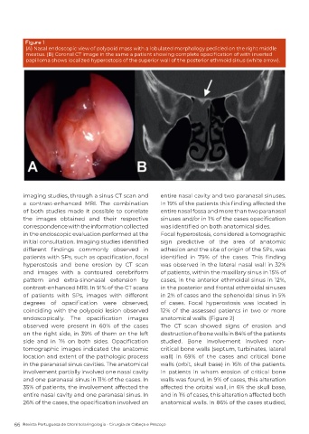
Figure 1
          (A) Nasal endoscopic view of polypoid mass with a lobulated morphology pedicled on the right middle
          meatus. (B) Coronal CT image in the same a patient showing complete opacification of with inverted
          papilloma shows localized hyperostosis of the superior wall of the posterior ethmoid sinus (white arrow).


          imaging studies, through a sinus CT scan and      entire nasal cavity and two paranasal sinuses.
          a contrast-enhanced MRI. The combination          In 19% of the patients this finding affected the
          of both studies made it possible to correlate     entire nasal fossa and more than two paranasal
          the images obtained and their respective          sinuses and/or in 1% of the cases opacification
          correspondence with the information collected     was identified on both anatomical sides.
          in the endoscopic evaluation performed at the     Focal hyperostosis, considered a tomographic
          initial consultation. Imaging studies identified   sign predictive of the area of anatomic
          different  findings  commonly  observed  in       adhesion and the site of origin of the SPs, was
          patients with SPs, such as opacification, focal   identified  in  79%  of  the  cases.  This  finding
          hyperostosis and bone erosion by CT scan          was observed in the lateral nasal wall in 32%
          and images with a contoured cerebriform           of patients, within the maxillary sinus in 15% of
          pattern and extra-sinonasal extension by          cases, in the anterior ethmoidal sinus in 12%,
          contrast-enhanced MRI. In 91% of the CT scans     in the posterior and frontal ethmoidal sinuses
          of patients with SPs, images with different       in 2% of cases and the sphenoidal sinus in 5%
          degrees  of  opacification  were  observed,       of cases. Focal hyperostosis was located in
          coinciding with the polypoid lesion observed      12% of the assessed patients in two or more
          endoscopically.  The  opacification  images       anatomical walls. (Figure 2)
          observed were present in 60% of the cases         The CT scan showed signs of erosion and
          on the right side, in 39% of them on the left     destruction of bone walls in 84% of the patients
          side  and  in  1%  on  both  sides.  Opacification   studied. Bone involvement involved non-
          tomographic images indicated the anatomic         critical bone walls (septum, turbinates, lateral
          location and extent of the pathologic process     wall) in 69% of the cases and critical bone
          in the paranasal sinus cavities. The anatomical   walls (orbit, skull base) in 16% of the patients.
          involvement partially involved one nasal cavity   In patients in whom erosion of critical bone
          and one paranasal sinus in 11% of the cases. In   walls was found, in 9% of cases, this alteration
          35% of patients, the involvement affected the     affected the orbital wall, in 6% the skull base,
          entire nasal cavity and one paranasal sinus. In   and in 1% of cases, this alteration affected both
          26% of the cases, the opacification involved an   anatomical walls. In 86% of the cases studied,


      66  Revista Portuguesa de Otorrinolaringologia - Cirurgia de Cabeça e Pescoço