
Page 69 - Revista Portuguesa - SPORL - Vol 62. Nº1
P. 69
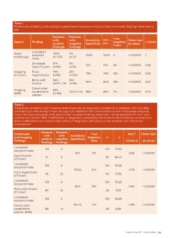
Table 1
          Prediction, reliability and statistical agreement measures of each of the commonly findings observed in
          SPs

                                    Patients   Patients
                                    with       with       Sensitivity  PV* +   True   Fisher test
          Search      Finding                                                negative             κ Cohen
                                    positive   negative   Specificity  PV* -          (p value)
                                    findings   findings                      Index
                      Lobulated
          Nasal       polypoid      100%       0%         100%        100%   0        < 0.00001   1
          endoscopy                 (n=103)    (n=0)
                      mass
                      Sinonasal     91%        9%         91%         91%    9%       < 0.00001   0.82
                      Opacification (n=91)     (n=9)
          Imaging     Focal         79%        21%
          (CT scan)   hyperostosis  (n=81)     (n=22)     79%         79%    12%      < 0.00001   0.62

                      Bony wall     84%        16%
                      erosion       (n=71 + 16)  (n=16)   84%         84%    16%      < 0.00001   0.67
                      Convoluted
          Imaging                   86%
          (MRI)       Cerebriform   (n=89)     14% (n=14)  86%        86%    7%       < 0.00001   0.73
                      pattern


          Table 2
          Prediction, reliability and influence level measures of diagnostic probability in patients with SPs after
          considering a clinical high index of suspicion related to the initial evidence of an endonasal polypoid
          mass. This was correlated with each of the imaging findings observed in these patients (CT scan and
          contrast-enhanced MRI). Coefficients of diagnostic probability estimates in all correlative comparisons
          show coefficients that reveal a high effect of diagnostic efficiency in the patients with SPs in our
          investigation

                            Patients  Patients
          Endoscopic                                          True                    Test T   Fisher test
          and imaging         with      with    Sensitivity  Negative   μ         σ
          findings           positive  negative  Specificity  Rate                   Cohen d     (p value)
                            findings  findings
          Lobulated
          polypoid mass        103        0                            51.1   72.83
                                                   91%         9%                      0,66     < 0.00001
          Opacification
          (CT scan)            91         9                             97    66.47
          Lobulated            103        0                            51.1   72.83
          polypoid mass
                                                  78.6%       21%                      0.78     < 0.00001
          Focal hyperstosis    81        22                             92    15.55
          (CT scan)
          Lobulated
          polypoid mass        103        0                            51.1   72.83
                                                   84%        16%                      0.84     < 0.00001
          Bony wall erosion    87        16                             95    11.31
          (CT scan)
          Lobulated            103        0                            51.1   72.83
          polypoid mass
          Convoluted                              86.4%       14%                      0.86     < 0.00001
          cerebriform          89        14                             96    9.89
          pattern (MRI)


                                                                                     Volume 62 . Nº1 . Março 2024 69