
Page 72 - Revista Portuguesa - SPORL - Vol 62. Nº1
P. 72
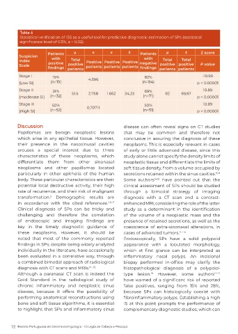
Table 6
          Statistical verification of ISS as a useful tool for predictive diagnostic estimation of SPs (statistical
          significance level of 0.5%, z = 0.02).

                        Patients    μ        σ        σ       x̄    Patients    μ        x̄      Z score
          Suspicion       with                                        with
          Index         positive   Total  Positive Positive Positive  negative  Total  Total
                                                                             positive positive
                                 positive
          Scale         findings  patients  patients patients patients  findings  patients patients  P value
          Stage I         19%              4.596                      82%                        -10.90
          (Low SI)       (n=19)                                      (n=84)                    p < 0.00001
          Stage II        31%      51.5    2.758    1.662   34,33     69%       82     69,67      10.89
          (moderate SI)  (n=32)                                      (n=71)                    p < 0.00001
          Stage III       50%                                         50%                         10.89
          (High SI)      (n=52)           0,70711                    (n=51)                    p < 0.00001


         Discussion                                         disease can often reveal signs on CT studies
         Papillomas are benign neoplastic lesions           that may be common and therefore not
         which arise in any epithelial tissue. However,     conclusive in assuring the diagnosis of these
         their presence in the nasosinusal cavities         neoplasms. This is especially relevant in cases
         arouses a special interest due to three            of early or little advanced disease, since this
         characteristics of these neoplasms, which          study alone cannot specify the density limits of
         differentiate them from other sinonasal            neoplastic tissue and differentiate the limits of
         neoplasms and other papillomas located             soft tissue density, from a volume occupied by
         particularly in other epithelia of the human       secretions retained within the sinus cavities. 11,13
         body. These particular characteristics are their   Some authors   14,15   have pointed out that the
         potential  local destructive  activity,  their  high   clinical assessment of SPs should be studied
         rate of recurrence, and their risk of malignant    through a bimodal strategy of imaging
         transformation.  Demographic results are           diagnosis with a CT scan and a contrast-
                         2
         in accordance with the cited references.   4,5–7    enhanced MRI, considering the role of the latter
         Clinical diagnosis of SPs can be tricky and        study  as  a  determinant  in  the  identification
         challenging and therefore the correlation          of  the  volume  of  a  neoplastic  mass  and  the
         of  endoscopic  and  imaging  findings  are        presence of retained secretions, as well as the
         key in the timely diagnostic guidance of           coexistence of extra-sinonasal alterations, in
         these neoplasms. However, it should be             cases of advanced tumors. 11 – 15
         noted that most of the commonly reported           Endoscopically, SPs have a solid polypoid
         findings in SPs, despite being widely analyzed     appearance with a lobulated morphology,
         individually in the literature, have occasionally   which  at  first  glance  can  be  interpreted  as
         been evaluated in a correlative way, through       inflammatory  nasal  polyps.  An  incisional
         a combined bimodal approach of radiological        biopsy  performed  in-office  may  clarify  the
         diagnosis with CT scans and MRIs.  8 – 12          histopathological  diagnosis  of  a  polypoid-
         Although a paranasal CT scan is indeed the         type lesion.  However, some authors        17–21
                                                                         16
         Gold Standard in the radiological study of         have warned of a significant risk of reported
         chronic  inflammatory  and  neoplastic  sinus      false positives, ranging from 15% and 28%,
         disease, because it offers the possibility of      because SPs can histologically coexist with
         performing anatomical reconstructions using        fibroinflammatory polyps. Establishing a high
         bone and soft tissue algorithms, it is essential   IS at this point prompts the performance of
         to highlight that SPs and inflammatory sinus       complementary diagnostic studies, which can


      72  Revista Portuguesa de Otorrinolaringologia - Cirurgia de Cabeça e Pescoço