
Page 67 - Revista Portuguesa - SPORL - Vol 62. Nº1
P. 67
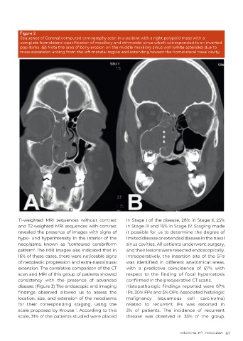
Figure 2
          Sequence of Coronal computed tomography scan in a patient with a right polypoid mass with a
          complete homolateral opacification of maxillary and ethmoidal sinus which corresponded to an inverted
          papilloma. (B) Note the area of bony erosion on the middle maxillary sinus wall (white asterisks) due to
          mass expansion arising from the left metatal region and extending toward the homolateral nasal cavity.


          T1-weighted MRI sequences without contrast        in Stage I of the disease, 28% in Stage II, 25%
          and T2-weighted MRI sequences with contrast       in Stage III and 16% in Stage IV. Staging made
          revealed the presence of images with signs of     it possible for us to determine the degree of
          hypo- and hyperintensity in the interior of the   limited disease or extended disease in the nasal
          neoplasms, known as "contoured cerebriform        sinus cavities. All patients underwent surgery,
          pattern". The MRI images also indicated that in   and their lesions were resected endoscopically.
          16% of these cases, there were noticeable signs   Intraoperatively, the insertion site of the SPs
          of neoplastic progression and extra-nasosinusal   was  identified  in  different  anatomical  areas,
          extension. The correlative comparison of the CT   with a predictive coincidence of 67% with
          scan and MRI of this group of patients showed     respect  to  the  finding  of  focal  hyperostosis
          consistency with the presence of advanced         confirmed in the preoperative CT scans.
          disease. (Figure 3) The endoscopic and imaging    Histopathologic  findings  reported  were  67%
          findings  observed  allowed  us  to  assess  the   IPs, 30% FPs and 3% OPs. Associated histologic
          location, size, and extension of the neoplasms    malignancy     (squamous    cell   carcinoma)
          for  their  corresponding  staging,  using  the   related to recurrent IPs was reported in
          scale proposed by Krouse  . According to this     3% of patients. The incidence of recurrent
                                    1
          scale, 31% of the patients studied were placed    disease  was observed in 33% of the group.


                                                                                     Volume 62 . Nº1 . Março 2024 67