Page 52 - Revista Portuguesa - SPORL - Vol 49. Nº3
P. 52

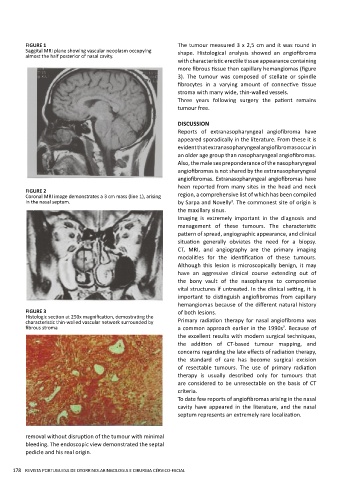
FIGURE 1                                          The tumour measured 3 x 2,5 cm and it was round in
          Saggital MRI plane showing vascular neoplasm occupying   shape.  Histological  analysis  showed  an  angiofibroma
          almost the half posterior of nasal cavity.
                                                            with characteristic erectile tissue appearance containing
                                                            more fibrous tissue than capillary hemangiomas (figure
                                                            3).  The  tumour  was  composed  of  stellate  or  spindle
                                                            fibrocytes  in  a  varying  amount  of  connective  tissue
                                                            stroma with many wide, thin-walled vessels.
                                                            Three  years  following  surgery  the  patient  remains
                                                            tumour free.

                                                            DISCUSSION
                                                            Reports  of  extranasopharyngeal  angiofibroma  have
                                                            appeared sporadically in the literature. From these it is
                                                            evident that extranasopharyngeal angiofibromas occur in
                                                            an older age group than nasopharyngeal angiofibromas.
                                                            Also, the male sex preponderance of the nasopharyngeal
                                                            angiofibromas is not shared by the extranasopharyngeal
                                                            angiofibromas. Extranasopharyngeal angiofíbromas have
                                                            heen reported from many sites in the head and neck
          FIGURE 2
          Coronal MRI image demonstrates a 3 cm mass (line 1), arising   region, a comprehensive list of which has been compiled
          in the nasal septum.                              by Sarpa and Novelly . The commonest site of origin is
                                                                              3
                                                            the maxillary sinus.
                                                            Imaging  is  extremely  important  in  the  diagnosis  and
                                                            management  of  these  tumours.  The  characteristic
                                                            pattern of spread, angiographic appearance, and clinical
                                                            situation  generally  obviates  the  need  for  a  biopsy.
                                                            CT,  MRI,  and  angiography  are  the  primary  imaging
                                                            modalities  for  the  identification  of  these  tumours.
                                                            Although  this  lesion  is  microscopically  benign,  it  may
                                                            have  an  aggressive  clinical  course  extending  out  of
                                                            the  bony  vault  of  the  nasopharynx  to  compromise
                                                            vital structures if untreated. In the clinical setting, it is
                                                            important  to  distinguish  angiofibromas  from  capillary
                                                            hemangiomas because of the different natural history
          FIGURE 3                                          of both lesions.
          Histologic section at 250x magnification, demostrating the
          characteristic thin-walled vascular network surrounded by   Primary  radiation  therapy  for  nasal  angiofibroma  was
          fibrous stroma                                    a common approach earlier in the 1990s . Because of
                                                                                               7
                                                            the excellent results with modern surgical techniques,
                                                            the  addition  of  CT-based  tumour  mapping,  and
                                                            concerns regarding the late effects of radiation therapy,
                                                            the  standard  of  care  has  become  surgical  excision
                                                            of  resectable  tumours.  The  use  of  primary  radiation
                                                            therapy  is  usually  described  only  for  tumours  that
                                                            are considered to be unresectable on the basis of CT
                                                            criteria.
                                                            To date few reports of angiofibromas arising in the nasal
                                                            cavity  have  appeared  in  the  literature,  and  the  nasal
                                                            septum represents an extremely rare localization.


          removal without disruption of the tumour with minimal
          bleeding. The endoscopic view demonstrated the septal
          pedicle and his real origin.


     178 REVISTA PORTUGUESA DE OTORRINOLARINGOLOGIA E CIRURGIA CÉRVICO-FACIAL
   47   48   49   50   51   52   53   54   55   56   57