


Page 91 - Revista Portuguesa - SPORL - Vol 60. Nº3
P. 91
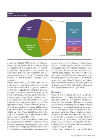
Figure 1
          Leucoplakia’s Histology


          presented a SCC; 68 (41,7%) had a premalignant    carcinoma was the histology on the first biopsy
          lesion and 45 (27,6%) had a benign lesion         (p=0,017), where higher grades of dysplasia
          as represented in Figure 1. For the analysis      were associated with higher progression rates.
          of  possible  risk  factors  for  progression  to   On the other hand, age, gender, tobacco or
          carcinoma, patients with malignant disease        alcohol  consumption,  smoking  cessation  or
          were excluded and only the 113 patients with      the presence of GERD weren’t associated with
          benign  or  pre-malignant  histology  were        the  progression  to  carcinoma.  Furthermore,
          analysed.                                         when  only  premalignant  lesions  are  taken
          Mean age was 58,67 ± 12,532 years and 93 (82,3%)   into consideration (n=68), the initial grade of
          patients were male. Leucoplakia was localised     dysplasia was still the only factor associated
          on the left  vocal fold in 37 (32,7%) patients,   with the progression to SCC (p=0,004).
          on the right vocal fold in 42 (37,2%) and was
          bilateral in 34 (30,1%). Furthermore, 84 (74,3%)   Discussion
          patients smoked, where 33 (29,2%) claimed to      Vocal  fold  leucoplakia  can  have  a  benign,
          have stopped smoking after the diagnosis, 17      premalignant or malignant histology with
          (15%) reported alcohol consumption, 16 (14,4%)    reports of up to 50% of the lesions being non-
          had a clinically diagnosed GERD and 6 (5,3%)      benign in the literature . This study included
                                                                                    4
          reported vocal abuse. Median follow-up was 24     163 patients submitted to MLS due to vocal fold
          (8 - 43,5) months. During this period 39 patients   leucoplakia in a tertiary hospital centre where
          had a new lesion and were submitted to an         31% of the lesions were malignant and 43%
          additional MLS, where 25 (22,1%) performed a      were pre-malignant. These results are higher
          total of 2 surgeries, 8 (7,1%) performed 3 and 6   than what has been reported previously . Thus,
                                                                                                   4
          (5,3%) were submitted to 4 MLS.                   patients with leucoplakia should be followed
          During follow-up 15 (13,3%) patients progressed   carefully and a biopsy should be performed
          to SCC. Comparison of different characteristics   since the percentage of malignant and pre-
          between patients that progressed to SCC and       malignant lesions could be higher than what
          patients that didn’t progress are listed on Table   was previously thought.
          1. The only factor associated with progression to   It has been established that vocal fold


                                                                                  Volume 60 . Nº3 . Setembro 2022 247

