
Page 96 - Revista Portuguesa - SPORL - Vol 50 Nº1
P. 96
were performed, showing air in the left cervical DISCUSSION
soft tissue; chest radiography showed no sing of Despite its frequency, tonsillectomy and adenoidectomy
pneumomediatinum or pneumothorax. Finally, a CT are rarely accompanied by perioperative complications.
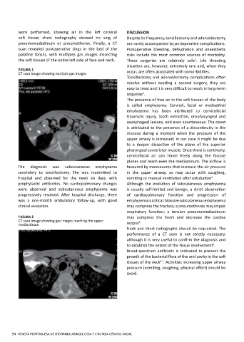
scan revealed postoperative sings in the bed of the Postoperative bleeding, dehydration and anaesthetic
palatine tonsils, with multiples gas images dissecting risks include the most common sources of morbidity.
the soft tissues of the entire left side of face and neck. These surgeries are relatively safe . Life threating
2
situation are, however, extremely rare and, when they
FIGURA 1 occur, are often associated with comorbidities.
CT scan image showing multiple gas images
Tonsillectomy and adenoidectomy complications often
resolve without needing a second surgery, they are
easy to treat and it is very difficult to result in long-term
3
sequelae .
The presence of free air in the soft tissues of the body
is called emphysema. Cervical, facial or mediastinal
emphysema has been attributed to cervicofacial
traumatic injury, tooth extraction, oropharyngeal and
oesophageal lesions, and even spontaneous. The onset
is attributed to the presence of a discontinuity in the
mucosa during a moment when the pressure of the
upper airway is increased; in our case it might be due
to a deeper dissection of the plane of the superior
pharyngeal constrictor muscle. Once there is continuity,
cervicofacial air can travel freely along the fasciae
planes and reach even the mediastinum. The airflow is
The diagnosis was subcutaneous emphysema favoured by manoeuvres that increase the air pressure
secondary to tonsillectomy. She was readmitted to in the upper airway, as may occur with coughing,
4
hospital and observed for the newt six days, with vomiting or manual ventilation after extubation .
prophylactic antibiotics. No cardiopulmonary changes Although the evolution of subcutaneous emphysema
were observed and subcutaneous emphysema was is usually self-limited and benign, a strict observation
progressively resolved. After hospital discharge, there of cardiopulmonary function and progression of
was a one-month ambulatory follow-up, with good emphysema is critical. Massive subcutaneous emphysema
clinical evolution. may compress the trachea; a pneumothorax may impair
respiratory function; a tension pneumomediastinum
FIGURA 2 may compress the heart and decrease the cardiac
CT scan image showing gas images reaching the upper 5
mediastinum output .
Neck and chest radiographs should be requested. The
performance of a CT scan is not strictly necessary,
although it is very useful to confirm the diagnosis and
6
to establish the extent of the tissue involvement .
Broad-spectrum antibiotic is indicated to prevent the
growth of the bacterial flora of the oral cavity in the soft
5,7
tissues of the neck . Activities increasing upper airway
pressure (vomiting, coughing, physical effort) should be
avoid.
94 REVISTA PORTUGUESA DE OTORRINOLARINGOLOGIA E CIRURGIA CÉRVICO-FACIAL