
Page 70 - Revista Portuguesa - SPORL - Vol 62. Nº2
P. 70
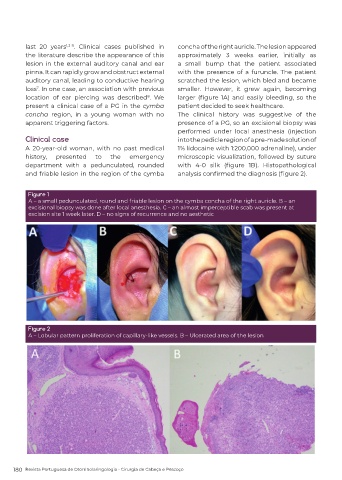
last 20 years 1,3-8 . Clinical cases published in   concha of the right auricle. The lesion appeared
         the literature describe the appearance of this     approximately 3 weeks earlier, initially as
         lesion in the external auditory canal and ear      a small bump that the patient associated
         pinna. It can rapidly grow and obstruct external   with the presence of a furuncle. The patient
         auditory canal, leading to conductive hearing      scratched the lesion, which bled and became
         loss . In one case, an association with previous   smaller.  However,  it grew again,  becoming
             7
         location of ear piercing was described . We        larger  (figure  1A)  and  easily  bleeding,  so  the
                                                  8
         present a clinical case of a PG in the cymba       patient decided to seek healthcare.
         concha region, in a young woman with no            The clinical history was suggestive of the
         apparent triggering factors.                       presence of a PG, so an excisional biopsy was
                                                            performed  under  local  anesthesia  (injection
         Clinical case                                      into the pedicle region of a pre-made solution of
         A 20-year-old woman, with no past medical          1% lidocaine with 1:200,000 adrenaline), under
         history,   presented    to   the   emergency       microscopic visualization, followed by suture
         department with a pedunculated, rounded            with  4-0  silk  (figure  1B).  Histopathological
         and friable lesion in the region of the cymba      analysis confirmed the diagnosis (figure 2).


          Figure 1
          A – a small pedunculated, round and friable lesion on the cymba concha of the right auricle. B – an
          excisional biopsy was done after local anesthesia. C – an almost imperceptible scab was present at
          excision site 1 week later. D – no signs of recurrence and no aesthetic


          Figure 2
          A – Lobular pattern proliferation of capillary-like vessels. B – Ulcerated area of the lesion


     180  Revista Portuguesa de Otorrinolaringologia - Cirurgia de Cabeça e Pescoço