
Page 92 - Portuguese Journal - SPORL - Vol 61. Nº2
P. 92
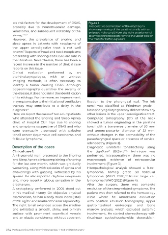
are risk factors for the development of OSAS,      Figure 1
          probably due to neuromuscular damage,              Preoperative examination of the oropharynx
                                                             shows asymmetry of the palatine tonsils, with an
          xerostomia, and subsequent instability of the      enlarged right tonsil. Note: the right anterior tonsil
          airway. 4,5,6,7                                    pillar was retracted posteriorly to the upper pole of
          However, the prevalence of snoring and             the tonsil for better exposure
          sleep apnea in patients with neoplasms of
          the upper aerodigestive tract is not well
          known. Reports of head and neck neoplasms
                 7
          presenting with snoring and OSAS are rare in
          the literature. Nevertheless, there has been a
          recent increase in the number of clinical case
          reports on this issue.
          Clinical  evaluation   performed     by    an
          otorhinolaryngologist,   with   or   without
          imaging methods,  is often necessary  to
          identify a tumor causing OSAS. Although
          polysomnography  quantifies  the  severity  of
          the disease, it does not aid in the identification
          of its etiology. Furthermore, an improvement
          in symptoms due to the initiation of ventilation   fixation  to  the  pharyngeal  wall.  The  left
          therapy may contribute to a delay in the          tonsil  was  classified  as  Friedman  grade  I.
          diagnosis. 8                                      Nasopharyngolaryngoscopy did not show any
          Here, we report the cases of two adult patients   other lesions in the upper aerodigestive tract.
          who attended the Snoring and Sleep Apnea          Computed tomography (CT) of the neck
          clinic of Hospital CUF Tejo due to snoring        showed a tumor originating in the palatine
          and symptoms suggestive of OSAS and who           tonsil with a transverse diameter of 30 mm
          were eventually diagnosed with palatine           and antero-posterior diameter of 22 mm,
          tonsil cancer (squamous cell carcinoma  and       without  changes  in  the  permeability  of  the
          follicular lymphoma).                             parapharyngeal space or presence of cervical
                                                            adenopathy (Figure 2).
          Description of the cases                          Diagnostic unilateral tonsillectomy using
          Clinical case 1:                                  the LigaSure   (BiZact )  technique  was
                                                                                     TM
                                                                          ®
          A 48 year-old man  presented to the Snoring       performed. Intraoperatively, there was no
          and Sleep Apnea clinic complaining of snoring     macroscopic     evidence   of   extracapsular
          for the last one month, which was gradually       involvement (Figure 3).
          worsening, along with episodes of apnea and       Histopathological  analysis  showed  a  B-cell
          awakenings with gasping, witnessed by his         lymphoma, namely grade 3B follicular
          spouse. He also reported daytime sleepiness       lymphoma  (WHO  2017)/follicular  large  cell
          and, more recently, globus sensation in the       lymphoma (WHO 2022) (Figure 4).
          oropharynx.                                       After the surgery, there was complete
          A  septoplasty  performed  in  2005  stood  out   resolution of the sleep-related symptoms. The
          in his medical history. On objective physical     patient was then referred to the hematology
          examination, he had a body mass index (BMI)       clinic where he underwent evaluation
          of 28.1 kg/m  and marked tonsillar asymmetry.     with  positron  emission  tomography,  upper
                     2
          The right tonsil extended across the midline      gastrointestinal   endoscopy,    and     bone
          and exhibited a smooth, shiny, and pinkish        marrow analysis, which excluded systemic
          surface  with  prominent  superficial  vessels    involvement. He started chemotherapy with
          and an elastic consistency, without apparent      rituximab, cyclophosphamide, doxorubicin,


     234  Portuguese Journal of Otorhinolaryngology - Head and Neck Surgery