
Page 100 - Portuguese Journal - SPORL - Vol 61. Nº2
P. 100
neck abscess and mediastinitis. As this is Description of the clinical case
a rare clinical condition, its presentation, A 49-year-old man was referred to the
assessment, and therapeutic approach have emergency department because of
not yet been established. 1 odynophagia, dysphagia, and pharyngeal
Here, we present a rare clinical case of late foreign body sensation for six weeks. He
pharyngeal perforation due to the extrusion denied other symptoms such as sialorrhea,
of an osteosynthesis screw 30 years after its dyspnea, hemoptysis, headache, fever, and
implantation in the cervical spine, as well as a paresthesia. He gave a previous history of
literature review on the topic. anterior cervical spine surgery in which
osteosynthesis of a fracture of the odontoid
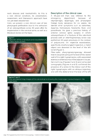
Figure 1 process with an interfragmentary screw was
Inspection of the oropharynx and visualization of performed 30 years previously in the context
a metal object (screw)
of polytrauma. On objective examination,
specifically oropharyngeal inspection, a metal
object was detected at the level of the left
tonsil (Figure 1).
Fiber-optic pharyngolaryngoscopy showed
that it was protruding at the level of the lateral
wall of the right hypopharynx, with associated
edema and hematoma of the adjacent tissues.
Cervical X-ray (Figures 2 and 3) and computed
tomography (Figures 4 and 5) confirmed the
presence of a screw in the lateral wall of the
right hypopharynx with an oblique trajectory
and with the distal end at the level of the left
Figures 2 and 3
Front and lateral X-ray images of the cervical spine showing the metal object (screw) lodged in the
oropharynx
242 Portuguese Journal of Otorhinolaryngology - Head and Neck Surgery