
Page 101 - Portuguese Journal - SPORL - Vol 61. Nº2
P. 101
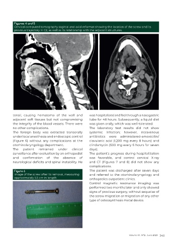
Figures 4 and 5
          Cervical computed tomography sagittal and axial reformat showing the location of the screw and its
          previous trajectory in C2, as well as its relationship with the adjacent structures.


          tonsil, causing hematoma of the wall and          was hospitalized and fed through a nasogastric
          adjacent soft tissues but not compromising        tube for 48 hours. Subsequently, a liquid diet
          the integrity of the blood vessels. There were    was given orally, which was well-tolerated.
          no other complications.                           The laboratory test results did not show
          The foreign body was extracted transorally        systemic infection; however, intravenous
          under local anesthesia and endoscopic control     antibiotics  were  administered-amoxicillin/
          (Figure  6)  without  any  complications  at  the   clavulanic acid (1,200 mg every 8 hours) and
          otorhinolaryngology department.                   clindamycin (600 mg every 6 hours for seven
          The   patient    remained    under     clinical   days).
          surveillance after evaluation by an orthopedist   The patient’s progress during hospitalization
          and   confirmation    of   the   absence   of     was favorable, and control cervical X-ray
          neurological deficits and spinal instability. He   and CT (Figures 7 and 8) did not show any
                                                            complications.
          Figure 6                                          The patient was discharged after seven days
          Image of the screw after its removal, measuring   and referred to the otorhinolaryngology and
          approximately 5.5 cm in length                    orthopedics outpatient clinics.
                                                            Control magnetic resonance imaging was
                                                            performed two months later and only showed
                                                            signs of previous surgery, without sequelae of
                                                            the screw migration or migration of any other
                                                            type of osteosynthesis metal device.


                                                                                      Volume 61 . Nº2 . June 2023 243