


Page 108 - Portuguese Journal - SPORL - Vol 61. Nº1
P. 108
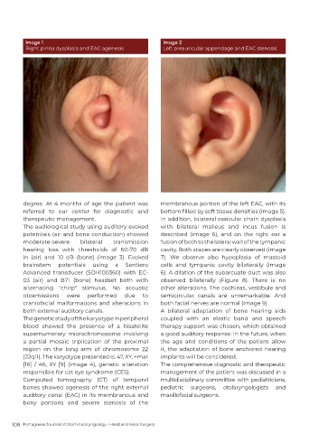
Image 1                                            Image 2
          Right pinna dysplasia and EAC agenesis             Left preauricular appendage and EAC stenosis


          degree. At 4 months of age the patient was        membranous portion of the left EAC, with its
          referred  to  our  center  for  diagnostic  and   bottom filled by soft tissue densities (image 5).
          therapeutic management.                           In addition, bilateral ossicular chain dysplasia
          The audiological study using auditory evoked      with  bilateral  malleus  and  incus  fusion  is
          potentials (air and bone conduction) showed       described  (image 6), and on the right ear a
          moderate-severe      bilateral   transmission     fusion of both to the lateral wall of the tympanic
          hearing loss with thresholds of 60-70 dB          cavity. Both stapes are clearly observed (image
          in (air) and 10 dB (bone) (image 3). Evoked       7). We observe also hypoplasia of mastoid
          brainstem   potentials   using   a   Sentiero     cells and tympanic cavity bilaterally (image
          Advanced  transducer  (SOH100360)  with  EC-      6). A dilation of the subarcuate duct was also
          03 (air) and B71 (bone) headset both with         observed bilaterally (Figure 8). There is no
          alternating "chirp" stimulus. No acoustic         other alterations. The cochleas, vestibule and
          otoemissions    were   performed     due   to     semicircular  canals  are  unremarkable.  And
          craniofacial malformations and alterations in     both facial nerves are normal (image 9)
          both external auditory canals.                    A bilateral adaptation of bone hearing aids
          The genetic study of the karyotype in peripheral   coupled with an elastic band and speech
          blood  showed  the  presence  of  a  bisatelite   therapy support was chosen, which obtained
          supernumerary microchromosome involving           a good auditory response. In the future, when
          a  partial  mosaic  triplication  of  the  proximal   the age and conditions of the patient allow
          region on the long arm of chromosome 22           it, the adaptation of bone anchored hearing
          (22q11). The karyotype presented is: 47, XY, +mar   implants will be considered.
          [16] / 46, XY [9] (image 4), genetic alteration   The comprehensive diagnostic and therapeutic
          responsible for cat eye syndrome (CES).           management of the patient was discussed in a
          Computed  tomography  (CT)  of  temporal          multidisciplinary committee with pediatricians,
          bones showed agenesis of the right external       pediatric  surgeons,   otolaryngologists  and
          auditory canal (EAC) in its membranous and        maxillofacial surgeons.
          bony portions and severe stenosis of the


     108  Portuguese Journal of Otorhinolaryngology - Head and Neck Surgery

