


Page 103 - Portuguese Journal - SPORL - Vol 61. Nº1
P. 103
clinical  improvement     and   the   patient     the infection among dogs (the main reservoir
          developed nephrotoxicity and muscle pain;         of human leishmaniasis) and cats is a cause
          therefore, amphotericin B was replaced by         for  concern  regarding  the  risk  of  increased
          itraconazole (600 mg once/day), which was         number of cases and dissemination of this
          given for three months. Subsequently, clinical    parasitosis. 5,6
          resolution  was  achieved  with  improvement      The severity of the clinical manifestations of
          in the erythema, nasal congestion, epistaxis,     leishmaniasis depends on factors such as the
          and nasal discomfort; in addition, the post-      parasite, the natural host’s resistance, and the
          treatment biopsies showed no Leishman-            magnitude of the immune response. Visceral
          Donovan bodies.                                   leishmaniasis  is  usually  more  severe  and  is
          The patient is being followed up in the           often fatal if not treated. HIV increases the
          dermatology and ORL clinics and, so far, has      host’s susceptibility to active disease, which
          exhibited no further manifestations  of the       usually manifests as a more aggressive and
          disease.                                          atypical form. In Portugal, there has been an
                                                            increase in the number of cases of visceral
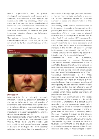
          Figure 3                                          leishmaniasis in adults with HIV co-infection,
          After treatment                                   a trend that  has  been decreasing  since the
                                                            introduction of anti-retroviral therapy. 2,9
                                                            Leishmaniasis      may      be     cutaneous,
                                                            mucocutaneous,      or  visceral.  Cutaneous
                                                            and  mucocutaneous  leishmaniasis  is not  a
                                                            notifiable  disease;  therefore,  it  is  believed  to
                                                            be underdiagnosed given the usually benign
                                                            course of the lesions. Visceral leishmaniasis
                                                            has been a notifiable disease since the 1950s.
                                                                                                         2
                                                            Cutaneous     leishmaniasis   is  the    most
                                                            common presentation of the disease and is
                                                            characterized by single or multiple pinkish
                                                            macular lesions that progress to nodules and
                                                            subsequently  to  painless  well-defined  ulcers
                                                            with raised borders that can affect any area of
                                                            the body. It is usually accompanied by painless
                                                            enlarged lymph nodes located near the site of
          Discussion                                        parasite inoculation. 1,3,8
          Leishmaniasis    is  a   primarily   zoonotic     Mucocutaneous  leishmaniasis  is  the  rarest
          infection, caused by protozoan parasites of       manifestation of the disease and is mainly
          the  genus  Leishmania  spp.  All  species  of    caused by L. Braziliensis. It is almost always
          Leishmania are transmitted through the bite       secondary to the cutaneous presentation.
          of female sand flies of the genera Lutzomyia      It may appear when the cutaneous lesion is
          and Phlebotomus. Transmission occurs by           still active, but it usually appears months or
          inoculation of the promastigote forms into the    years after its resolution. It primarily affects
          skin of the vertebrate host. 1,3,8                the nasal mucosa; however, as the disease
          This disease is mostly endemic to the tropical    becomes more aggressive, it can affect the
          rural areas of Central and South America (New     mucosa of the oral cavity, pharynx, and larynx.
          World),  Africa,  India,  and  the  Middle  East   The anterior region of the nasal septum is a
          (Old World), as well as Mediterranean regions     favorable environment for the development
          of Europe. In Portugal, the most recently         of amastigotes. Low temperatures in this part
          collected data show that the prevalence of        decrease the ability of macrophages to carry


                                                                                     Volume 61 . Nº1 . March 2023 103

