


Page 110 - Portuguese Journal - SPORL - Vol 61. Nº1
P. 110
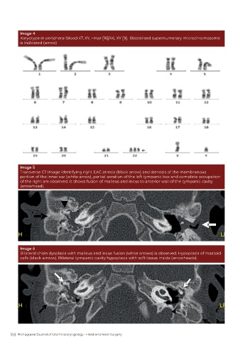
Image 4
          Karyotype in peripheral blood 47, XY, +mar [16]/46, XY [9].  Bisatelised supernumerary microchromosome
          is indicated (arrow)


          Image 5
          Transverse CT image identifying right EAC atresia (black arrow) and stenosis of the membranous
          portion of the inner ear (white arrow), partial aeration of the left tympanic box and complete occupation
          of the right are observed. It shows fusion of malleus and incus to anterior wall of the tympanic cavity
          (arrowhead).


          Image 6
          Bilateral chain dysplasia with malleus and incus fusion (white arrows) is observed. Hypoplasia of mastoid
          cells (black arrows). Bilateral tympanic cavity hypoplasia with soft tissue inside (arrowheads).


      110  Portuguese Journal of Otorhinolaryngology - Head and Neck Surgery

