


Page 102 - Portuguese Journal - SPORL - Vol 61. Nº1
P. 102
cutaneous,  mucocutaneous, and visceral.          of the septum. Concomitantly, an anterior
          Mucocutaneous  leishmaniasis  is  the  rarest     septal  perforation  of  approximately  0.50.5
          form and is mainly caused by the species          cm was observed in the Cottle areas 2 and 3
          Leishmania Braziliensis. It may occur as          (Figure 2).  Neck palpation was normal, as was
                                                                      7
          a primary manifestation of the disease or         the remaining objective ORL examination.
          secondary to the cutaneous presentation. 1,3,6    There were no erythematous lesions at other
          The case presented herein is that of a            sites of the body.
          woman with a previous history of cutaneous         Figure 2
          leishmaniasis treated five years ago who was       Nasal endoscopy of the right nasal cavity. Multiple
          referred to the otorhinolaryngology (ORL)          small lesions, with erythematous borders can be
          clinic due to de novo nasal complaints.            seen


          Case description
          The patient was a 48-year-old woman who
          lived in a rural area and was referred to the
          ORL clinic for nasal congestion and self-
          limited episodes of epistaxis. She had a
          relevant  history  of  type  II  diabetes  mellitus
          and cutaneous leishmaniasis (Leishmania
          tropica) diagnosed in 2015, which was treated
          with oral itraconazole (600 mg once/day for
          28 days), with lesion resolution. She did not
          have a history of nasal trauma or chronic
          rhinosinusitis. She denied having recently
          travelled abroad or before the diagnosis of
          cutaneous leishmaniasis.
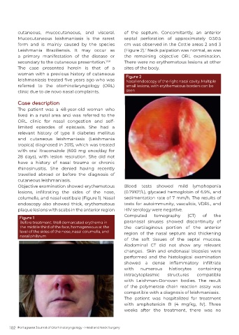
          Objective examination showed erythematous         Blood  tests  showed  mild  lymphopenia
          lesions,  infiltrating  the  sides  of  the  nose,   (0.7910 /L), glycated hemoglobin of 6.9%, and
                                                                   9
          columella, and nasal vestibule (Figure 1). Nasal   sedimentation rate of 7 mm/h. The results of
          endoscopy  also  showed  thick,  erythematous     tests for autoimmunity, vasculitis, VDRL, and
          plaque lesions with scabs in the anterior region   HIV serology were negative.
                                                            Computed      tomography      (CT)   of    the
          Figure 1
          Before treatment. Well demarcated erythema in     paranasal sinuses showed discontinuity of
          the middle third of the face, homogeneous at the   the cartilaginous portion of the anterior
          level of the sides of the nose, nasal columella, and   region of the nasal septum and thickening
          nasal philtrum
                                                            of the soft tissues of the septal mucosa.
                                                            Abdominal CT did not show any relevant
                                                            changes.  Skin and endonasal biopsies were
                                                            performed  and  the  histological  examination
                                                            showed  a  dense  inflammatory  infiltrate
                                                            with    numerous     histiocytes   containing
                                                            intracytoplasmic    structures    compatible
                                                            with  Leishman-Donovan  bodies.  The  result
                                                            of  the  polymerase  chain  reaction  assay  was
                                                            compatible with a diagnosis of leishmaniasis.
                                                            The  patient was  hospitalized for  treatment
                                                            with amphotericin B (4 mg/kg, IV). Three
                                                            weeks after the treatment, there was no


      102  Portuguese Journal of Otorhinolaryngology - Head and Neck Surgery

