
Page 9 - Revista Portuguesa - SPORL - Vol 61. Nº4
P. 9
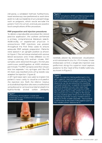
(48 pens), a validated method. Furthermore,        Figure 2
          nasal endoscopy was performed at each time         Platelet rich plasma (PRP) prefilled syringes after
                                                             centrifugation (one for each nasal cavity).
          point to rule out baseline structural pathology
          such as polyposis, which would exclude the
          patient from the cohort, and evaluate possible
          local complications of the procedure.

          PRP preparation and injection procedures
          To obtain a reproducible procedure for clinical
          practice application, the authors performed
          a primary comprehensive literature search.
          Additionally, an experienced physician in
          osteoarticular PRP injections was present
          throughout  the  first  three  cases  to  ensure
          adequate PRP sample preparation. Patients
          were seated in an upright position as shown
          in Figure 1. The technique started with venous
          blood  extraction  onto  three  different  3.5  ml   carefully placed by endoscopic visualization
          tubes containing 3.2% sodium citrate. PRP         and maintained in situ for ≥ 10 minutes. Under
          samples were obtained through a 10-minutes        endoscopic control, a single-site injection was
          continuous centrifugation at 4000 rotations       performed along the superior  nasal septum
          per minute. The PRP samples were then drawn       posterior to the head of the middle turbinate,
          into two separate 1‐mL syringes until the 0.9     as shown in Figure 3.
          ml mark was reached and a 27‐g needle was
          adapted for injection (Figure 2).                  Figure 3
          A 30º rigid nasal optic was used to inspect the    Single-site injection being performed in the
          patient’s nasal cavity to visualize and predict    right superior nasal septum, posteriorly to the
          the injection site. Both the inferior meatus       level of the head of the middle turbinate. PRP
                                                             dispersion can be perceived by the resulting
          and predicted injection site were anesthetized     mucosal blanching in a superior and posterior
          using lidocaine + prilocaine and phenylephrine     direction (dashed circular line). Care must
          hydrochloride    eluted   cotton    pledgets,      be taken to avoid pinching the presumable
                                                             site of S-point (blue circle), which can occur if
                                                             the puncture occurs too highly in the septal
           Figure 1
           Positioning of the patient´s nose and             mucosa. Dashed green lines and numeration
           endoscopy screen relative to the operator: a      are displaying three virtually marked vertical
                                                             divisions of the middle turbinate – the injection
           schematic model.
                                                             is performed at the level of the superior part of
                                                             the middle portion (labeled as 2).


                                                                                  Volume 61 . Nº4 . Dezembro 2023 347